


In this tutorial, we have explained the root anatomy along with the monocot root cross section and the dicot root cross section. Additionally, we did a difference between monocot vs dicot root.
TABLE OF CONTENTS
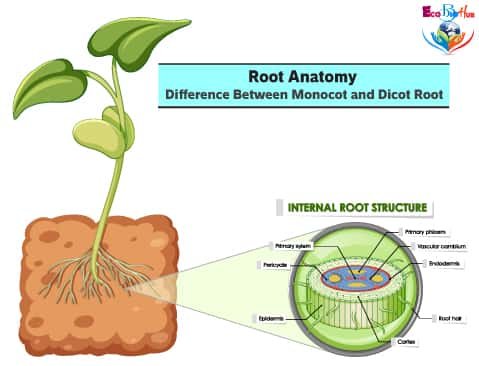
ROOT ANATOMY: DICOT ROOT CROSS SECTION
■ Dicot Root Diagram Reveals Internal Structure of Dicot Root

A thin transverse section of the young dicot root of Gram, Sunflower or Pea reveals the following structures under the microscope:
1. Epiblema or Piliferous Layer
This is the outermost layer of root-derived from the protoderm of the apical meristem. It consists of thin-walled parenchyma cells, the outer walls of which are uncutinised. The outer walls of most of the cells extend out into tubular and unicellular structures called root hair. Epiblema along with root hair helps in the absorption of water and mineral salts from the soil.
2. Cortex
It forms a major part of the root body and consists of several layers of oval or rounded thin-walled parenchyma cells. These cells contain starch grains and are loosely packed with intercellular spaces between them.
3. Endodermis
This is the innermost layer of the cortex consisting of barrel-shaped cells without intercellular spaces. The radial and the inner tangential walls of the cells of the endodermis are greatly thickened. In some cases, thickened strips of lignified material, called the Casparian strips run along the inner side of the radial and tangential walls. These strips are impervious to water. In most cases, certain cells of the endodermis lying against the protoxylem have thickenings only on their radial walls. The outer and inner tangential walls of these cells remain thin. Such cells are known as passage cells
4. Pericycle
This is a single layer of thin-walled parenchyma cells lying immediately below the endodermis. It gives protection to the vascular tissue.
5. Vascular Bundles
The vascular tissue occurs as an equal number of separate xylem and phloem bundles arranged in a ring on alternate radii. They are usually 2-4 in number. Such vascular bundles are called radial.
In the young root, cambium is absent. It makes its appearance afterwards. The xylem bundle consists of protoxylem and metaxylem. The protoxylem lies towards the periphery and the metaxylem towards the centre of the root. This arrangement of xylem is called exarch
6. Conjunctive Tissue
It is a mass of parenchyma cells lying between xylem and phloem bundles.
7. Pith
It is very small and parenchymatous. It is generally found in monocot roots.
■ Anatomical Characteristics of Dicot Roots
1. Xylem bundles two to four in number and may be diarch, triarch and tetrarch.
2. Pericycle gives rise to lateral roots and secondary meristems such as cambium and phellogen.
3. Cambium develops later on as secondary meristem.
4. Pith is usually absent.
ROOT ANATOMY: MONOCOT ROOT CROSS SECTION
■ Monocot Root Diagram Reveals Internal Structure of Monocot Root

The general structure of the monocot root of Zea mays is on the same pattern as of the dicot root. A transverse section of the root shows the following structures:
1. Epiblema
It has the same general structure and functions as in dicot root.
2. Cortex
It has the same general structure and functions as in dicot root except that the cortex is more extensive.
3. Endodermis
It has the same general structure and functions as in dicot root.
4. Pericycle
It is a single-cell layer lying internal to the endodermis. It consists of small, thin-walled parenchyma cells with abundant protoplasm.
5. Conjunctive Tissue
It is a mass of parenchyma cells lying in between the xylem and phloem bundles.
6. Pith
It occupies the central portion of the root and consists of parenchymatous cells. Unlike dicot roots, it is well-developed in monocot roots.
7. Vascular Bundles
The vascular tissue in monocot roots is polyarch. It is organized into 8 or more separate xylem and phloem bundles arranged in separate radial in an alternate fashion. Each xylem bundle consists of a protoxylem lying towards the pericycle and a metaxylem
■ Anatomical Characteristics of Monocot Roots
1. Cortex is massive with thin-walled parenchymatous cells with intercellular spaces.
2. Vascular bundles are more numerous, usually 8 or more in number.
3. Pericycle is partly parenchymatous and partly sclerenchymatous.
4. Pericycle gives rise to lateral roots.
5. Since cambium is absent, there is no secondary growth in roots.
6. Pith is large and well-developed.
MONOCOT vs DICOT ROOT
Characters
Monocot Root
Dicot Root
1. Vascular bundles
Polyarch i.e., more than 6 vascular bundles.
Diarch to hexarch i.e., vascular bundles varies from 2 to 6.
2. Cambium
Absent.
Present, so secondary growth occurs.
3. Xylem vessels
Xylem vessels are large and more or less circular in outline.
Xylem vessels are smaller in size and are polygonal in shape.
4. Cortex
Cortex is very wide.
Cortex is comparatively narrow.
5. Pith
Well developed large pith.
It is small and poorly developed or absent.
6. Activity of Pericycle
Pericycle gives rise to lateral roots only.
It gives rise to lateral roots, cork cambium and part of the vascular cambium.
7. Endodermis
Casparian strips are visible only in the young root. The endodermal cells later become highly thickened and give U-shaped appearance.
Endodermis is less thickened and Casparian strips are more prominent.
8. Conjunctive tissue
Parenchymatous or sclerenchymatous.
Parenchymatous
9. Secondary growth
Secondary growth is absent.
It develops after the appearance of cambium.
DICOT STEM vs DICOT ROOT
Sl. No.
Dicot Stem
Dicot Root
1. Outer Layer
The outermost layer of cells is known as epidermis.
The outermost layer of cells is called epiblema.
2. Cuticle
Epidermis is covered with a thin layer of cuticle.
Epiblema is not cutinised.
3. Activity of hair
In many cases, the epidermis bears multicellular hair which is simply protective.
Epiblema bears numerous unicellular hair. They help in the absorption of water and minerals from the soil.
4. Cortex
Cortex is narrower than in the root.
Cortex is wider.
5. Pericycle
Pericycle is made up of sclerenchymatous patches and the intervening masses of parenchyma. It lies in between the vascular bundles and the endodermis.
It consists of a single layer of thin-walled parenchyma cells.
6. Vascular bundles
Vascular bundles are conjoint and collateral or bicollateral.
They are arranged in a ring but are radial, i.e., phloem and xylem are found on different radii.
7. Xylem
Endarch.
Exarch.
8. Endodermis
Absent or poorly developed.
Well developed.
9. Ground tissue
A few outer cells of the ground tissues may contain chloroplasts.
Chloroplasts are almost invariably absent.