


In this tutorial, we have discussed the leaf anatomy (Transverse Section of Leaf) of dicot and monocot plants.
TABLE OF CONTENTS
Transverse Section of Leaf: Dorsiventral/ Dicotyledonous Leaf
The leaves of dicotyledonous plants are arranged in a horizontal position, i.e., at a right angle to the rays of the sun so that they receive more light on the upper surface than the lower surface. The leaves with such an arrangement are known as dorsiventral or bifacial leaves. This unequal illumination in these leaves is marked by the high degree of differentiation in the internal structure.
A thin section at the right angle to the surface of the dicot leaf, passing through the midrib, shows the following structures:

1. Upper Epidermis (Adaxial Epidermis)
It consists of a single layer of parenchymatous cells. The outer walls of the cells are covered with a layer of cuticle. Epidermal cells do not contain chloroplasts and epidermis.
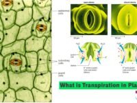
2. Lower Epidermis (Abaxial Epidermis)
This is also a single layer of cells on the lower surface of the leaf. Cuticle is absent or if present then comparatively thinner. The lower epidermis contains numerous stomata. Each stoma consists of a minute opening leading into a large air cavity. Stomata help in gaseous exchange between the atmosphere and the leaf tissues and also help in the evaporation of excess water from the leaf.
3. Mesophyll
The tissue in between the upper and lower epidermis constitutes mesophyll. It consists of parenchymatous cells and forms the major portion of the interior of the leaf. It is differentiated into:
(i) Palisade Parenchyma:
It lies just below the epidermis and consists of one or more layers of closely packed elongated cells with a few or no intercellular spaces. These cells lie with their long axis at the right angle to the epidermis. Palisade cells contain a large number of chloroplasts. The function of the palisade tissue is photosynthesis in the presence of sunlight.
(ii) Spongy Parenchyma:
It lies towards the lower epidermis and consists of round, oval, or irregular and thin-walled parenchymatous cells. These cells are loosely packed and have large intercellular spaces and air cavities. These contain a few chloroplasts. The mesophyll forms a compact mass of cells around the veins or bundles. Spongy parenchyma helps in the exchange of gases and water vapours through the intercellular spaces and air cavities which are in direct communication with the outer atmosphere through the stomata. These also help to some extent in the manufacture of food.
4. Vascular Bundles
Each vascular bundle consists of xylem and phloem.
Xylem lies towards the upper epidermis and consists of vessels, tracheids, xylem parenchyma, and wood fibres.
Phloem lies towards the lower epidermis and consists of sieve tubes, companion cells, and phloem parenchyma.
Xylem helps in the conduction and distribution of water and mineral salts to the leaf. Phloem translocates manufactured food from the leaf to the storage organs and other regions of the plant body.
The vascular bundles are surrounded partially or completely by a layer of thick-walled parenchymatous cells known as bundle sheath.
Transverse Section of Leaf: Monocotyledonous/ Isobilateral Leaf
A typical monocot leaf (Maize) in the transverse section shows the following structues:

1. Epidermis
There is no difference in the upper and the lower epidermis. Stomata occur on both surfaces and are surrounded by dumb-bell-shaped guard cells. Leaves with stomata on both surfaces are called amphistomatic leaves. The epidermis is made of compact cells which lack chloroplasts. In the upper epidermis, some cells become large-sized and are called bulliform or motor cells. These cells have large vacuoles containing water. These are supposed to play a role in rolling and unrolling of the mature leaves. According to another view, they are supposed to help in unrolling of leaf during its development.
2. Mesophyll
It is not differentiated into palisade and spongy parenchyma. These cells are almost spherical with small inter cellular spaces. These cells contain chloroplasts.
3. Vascular Bundles
In monocot leaves, vascular bundles occur as a large number of closely placed small and a few large vascular bundles. These are conjoint and collateral. Because of parallel venation, each vein comprises one vascular bundle. Each vascular bundle consists of a xylem towards the upper surface and phloem towards the lower surface. Vascular bundles are surrounded by a bundle sheath formed of a single layer of parenchymatous cells having chloroplasts.
The bundle sheath is of systematic importance in grasses. It is called panicoid when the sheath is single-layered, and festucoid when it is double-layered. The larger vascular bundles are surrounded by sclerenchymatous bundle sheath which may show bundle sheath extensions. Its cells contain chloroplasts.
The peculiar arrangement of tissues in a Maize leaf having bulliform cells in the upper epidermis and the undifferentiated mesophyll occurring in concentric layers around the enlarged bundle sheath cells with chloroplasts is known as Kranz’s anatomy.
Difference Between Monocot and Dicot Leaf
(i) Palisade parenchyma. (ii) Spongy parenchyma with large intercellular spaces.
Characters
Dicot / Dorsiventral Leaf
Monocot / Isobilateral Leaf
Symmetry
Dorsiventral. It is exposed horizontally to the sun rays, i.e., one side is exposed to the sunlight.
Isobilateral. Its both sides are equally exposed to sunlight.
Leaf surface
It has distinct upper and lower surfaces. The upper layer is more thickly cutinised.
Its both the surfaces are alike and equally cutinised.
Stomata distribution
Hypostomatic i.e., stomata are mostly present on the lower surface of leaf.
Amphistomatic. Stomata are equally distributed on both surfaces.
Mesophyll
Due to unequal illumination, mesophyll is differentiated into
Owing to equal illumination, mesophyll is not differentiated into palisade and spongy parenchyma. Only spongy parenchyma is present which has small intercellular spaces.
Bulliform cells
Present on upper epidermis.
Usually absent.
Vascular bundles
Vascular bundles have parenchymatous patches on both sides up to the epidermis.
Vascular bundles have sclerenchymatous patches on both sides up to the epidermis.
Vascular bundles position
Vascular bundles have downward phloem and upward xylem.
Vascular bundles are alike that of a monocot system, i.e., phloem on the upper side and xylem on the lower side.