


In this tutorial, we have described Stem Anatomy (Monocot and Dicot Stem Cross Section).
TABLE OF CONTENTS
DICOT STEM CROSS SECTION
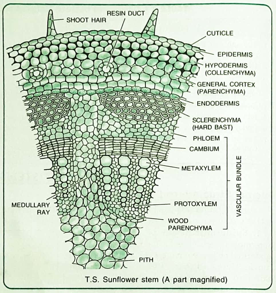
For studying the internal structure of a typical dicot stem, the stem cross section of a young Sunflower or Cucurbita is taken. A thin transverse section of the stem reveals the following structures under the microscope:

1. Epidermis
It forms a single and the outermost layer of the stem. The outer walls of the epidermal cells are covered with a thin layer of cuticle. Here and there, it bears multicellular hair and a few stomata.
The cells of the epidermis are all alike and parenchymatous.
It is protective in nature.
2. Cortex:
It lies below the epidermis and is many-layered. Cortex occupies the position between epidermis and pericycle, and is differentiated into the following three zones:
(i) Hypodermis:
It is a 4 to 5 cell-thick layer of collenchymatous cells which lies immediately below the epidermis. The collenchymatous cells are living and contain chloroplasts.
(ii) General Cortex:
It lies below the hypodermis. It consists of a few layers of thin-walled parenchymatous cells with conspicuous intercellular spaces. It is reduced to 1-2 layers outside the vascular bundles. Some of the cells have chloroplasts. Some resin ducts (oil ducts in Sunflower) are also seen which are surrounded by a ring of glandular cells.
It stores food and other materials. It also provides rigidity to the stem.
(iii) Endodermis:
It is the cortex innermost layer of the cortex. It is made up of a single row of
3. Pericycle
It is formed of semilunar patches of sclerenchyma above the phloem. It is 4-5 layers thick. The sclerenchyma cells are dead and rigid with their walls considerably thickened and lignified. Since these patches lie outside the phloem, they are known as hard bast.
Pericycle provides mechanical support to the plant and protects vascular bundles. It also stores food.
4. Vascular Bundles
In a dicot stem, a large number of vascular bundles are arranged in a ring enclosed by pericycle. Vascular bundles are conjoint, open and collateral and with endarch protoxylem.
In between the vascular bundles, there are a few layers of radially placed parenchymatous cells. They constitute medullary rays. In Cucurbita, the vascular bundles are bicollateral.
Each vascular bundle is composed of phloem, xylem and cambium.
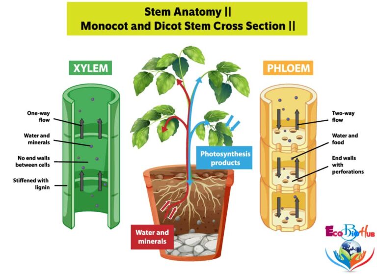
(i) Xylem:
It is the innermost layer of vascular bundles and lies towards the centre of the stem. Xylem consists of vessels, tracheids, wood fibres and wood parenchyma. The smaller vessels which lie towards the centre comprise of protoxylem and the bigger ones that lie away from the centre are known as metaxylem. Such xylem is known as endarch.
(ii) Phloem:
It lies towards the outer side and is composed of sieve tubes, companion cells and phloem parenchyma. The phloem cells are living and have thin walls of cellulose.
(iii) Cambium:
It is a strip of thin-walled cells lying in between phloem and xylem. The cambial cells are small, rectangular, thin-walled and radially arranged.
5. Medullary Rays or Pith Rays
These constitute radial rows of big and rectangular parenchymatous cells. Medullary rays lie in between vascular bundles and serve in the conduction of water and food in the radial direction.
6. Pith or Medulla
It is the central parenchymatous zone and in sunflower occupies the major portion of the stem. The parenchymatous cells are living, thin-walled and rectangular with conspicuous intercellular spaces.
MONOCOT STEM CROSS SECTION
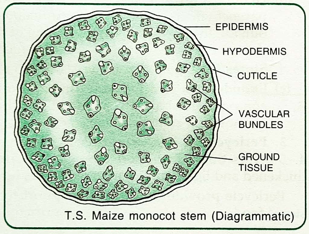
A thin transverse section through a monocot stem (Maize) reveals the following structures under the microscope:
1. Epidermis
It forms a single and the outermost layer of the stem. The epidermis consists of small barrel-shaped cells lying attached closely by the radial walls, the outer walls of which are cuticularised. Some stomata are also present.
Epidermis is protective in nature.
2. Hypodermis
It lies immediately below the epidermis. It consists of two or three layers of sclerenchymatous cells.
It provides mechanical support and strength to the stem.
3. Ground Tissue
It consists of a mass of thin-walled parenchymatous cells extending from below the hypodermis to the centre of stem.
It is not differentiated into definite tissues like cortex, endodermis, pericycle, etc., as in dicot stems. The cells of ground tissue have intercellular spaces.

4. Vascular Bundles
The vascular bundles are more numerous than in dicot stems and lie scattered in the ground tissue. They lie closer to the periphery than in the centre. The peripheral vascular bundles are smaller than the central ones.
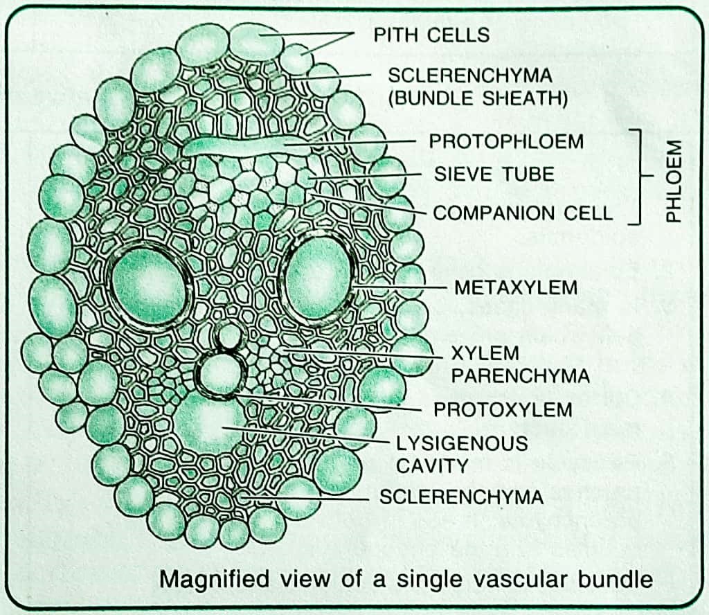
Each vascular bundle is surrounded by a sheath of thick-walled sclerenchymatous cells called the bundle sheath. It provides protection and strength to vascular bundles. The structure of the individual vascular bundle varies greatly in monocots. In Maize, the bundles are oval, conjoint, collateral
Since there is no cambium, monocots do not show any secondary growth. Each vascular bundle consists of xylem and phloem.
(i) Xylem:
It consists usually of four distinct vessels arranged in the form of ‘Y’. The two larger and rounded vessels situated on the sides with pitted tracheids in between them constitute metaxylem. The protoxylem consists of two smaller vessels situated towards the centre. Vessels of metaxylem are pitted and those of protoxylem are annular and spiral.
The thin-walled cells of wood parenchyma along with the sclerenchyma or wood fibres are seen lying in association with protoxylem On the inner side of xylem, a prominent water containing cavity or lysigenous cavity is seen. It is formed by the disorganisation of one or more protoxylem vessels in the parenchyma cells.
(ii) Phloem:
The phloem lies in between the arms of ‘Y’ facing the epidermis and consists exclusively of sieve tubes and companion cells.
Phloem parenchyma is absent. The protophloem which is the broken mass of the cells lies on the outer side and the inner portion constitutes the metaphloem.
DIFFERENCE BETWEEN MONOCOT AND DICOT STEM
(ii) Conjoint, collateral or bicollateral, endarch and open. (iii) Bundle sheath absent. (iv) Vascular bundles are of uniform size and wedge-shaped. (ii) Conjoint, collateral, endarch and closed. (iii) Sclerenchymatous bundle sheath is usually present. (iv) They are smaller near the periphery and bigger in the centre, and are oval-shaped. (ii) There is no lysigenous cavity in the xylem. (ii) Presence of lysigenous cavity in the xylem
Characters
Dicot Stem
Monocot Stem
Epidermis
The epidermis is single layered with multicellular hair and trichomes.
Single layered, without epidermal hair and trichomes are absent.
Hypodermis
Hypodermis is collenchymatous.
It is sclerenchymatous.
Ground tissue
Ground tissue is differentiated into cortex, endodermis, pericycle, pith etc.
Ground tissue is not differentiated into cortex, endodermis and pericycle, instead it consists of a mass of uniform and alike cells.
Endodermis
Present with starch grains, Casparian strips are generally absent.
Absent.
Pericycle
Few layered thick.
Absent.
Vascular bundles
(i) Vascular bundles are fewer in number and arranged in a ring.
(i) Numerous and lie scattered in the ground tissue.
Xylem vessels
(i) Xylem vessels are numerous and arranged in radial rows. They are polygonal in shape.
(i) Xylem vessels are a few in number and arranged in the form of ‘Y’ or ‘V’ and are oval or rounded in shape.
Phloem
Phloem consists of sieve tubes, companion cells and phloem parenchyma.
Phloem consists exclusively of sieve tubes and companion cells.
Medullary rays
Parenchymatous, found in between vascular bundles.
Absent.
Pith
Centrally placed with parenchymatous or occasionally sclerenchymatous cells.
Absent.
Phloem parenchyma
Present.
Absent.
Secondary growth
Secondary growth takes place due to the presence of cambium.
Secondary growth usually absent due to the absence of cambium.